Waggie – A Tumour In A Boxer
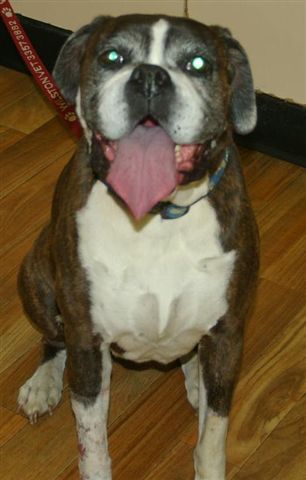
Name: Waggie
Species: Canine
Breed: Boxer
Gender: Female Speyed
Age: 9yrs
Weight: 27.0 kgs
History: Waggie was initially presented at Wilston Vet Surgery with lameness and a head tilt, however during examination Dr Meredith Brothers was also told that she had a lump on her leg. After examining and treating the initial health concerns, more attention was paid to this mysterious lump that Waggle had. It was about 2cm in diameter and was located on the outside of her right carpus (lower leg).
Examination: A FNA (fine needle aspirate ), which is where a needle is inserted into the lump to collect some cells, was done to try and determine what the lump was. This sample was sent off to the lab for analysis. The next day the results were back and they revealed the lump was a soft tissue sarcoma, which is a very aggressive type of cancer.
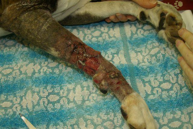
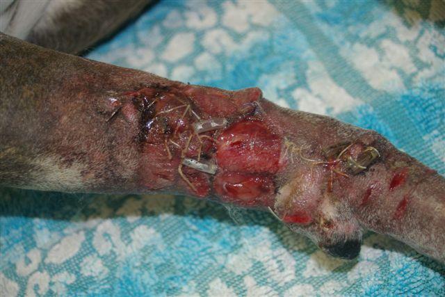
The Plan: This lump needed to be removed as soon as possible so Waggie was admitted to the hospital a few days later. Because this lump was cancerous, a large amount of tissue around it would need to be excised to lessen the chance of some cancer cells being left behind and the lump returning. Unfortunately this meant the wound would be very hard to close, so Waggie would need many trips to the clinic for bandage changes and wound management.
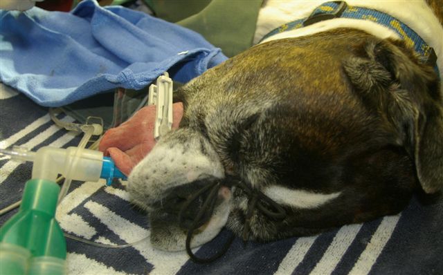
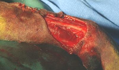
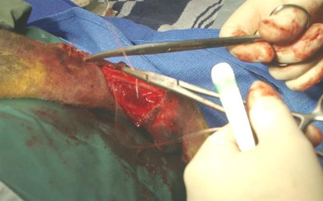
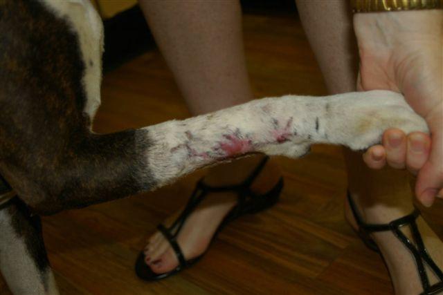
During surgery an incision of 1 ½ cm in diameter was made around the tumor, and a very deep margin that went under the flexor tendon and very close to the bone. After the tumor was removed, a 5cm open surgical wound remained. It was flushed with sterile saline then the attempt to close began. Dr Brothers brought together as much of the tissue as possible and made some tension cuts around the surgical wound to relieve some pressure on the skin. 3cm that was unable to be closed was left open to heal and granulate on its own.
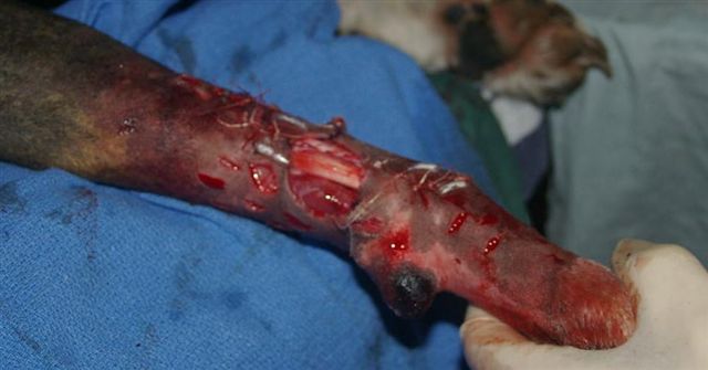
The first bandage was applied; this consisted of a contact layer of paraffin infused gauze, which is designed to not stick to the wound and provide a moist environment. Next a soft cotton wool layer is applied for comfort and padding. A layer of conforming bandage is wrapped over the soft layer, and lastly a stretchy, sticky layer called “vetwrap” goes on for protection of the inner layers and to hold everything in place.
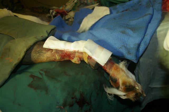
Waggie was sent home with oral antibiotics and pain killers as well as strict instructions to rest.
Week one:
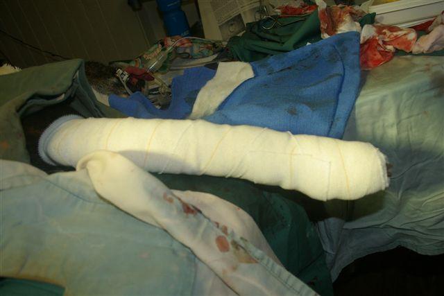
The wound is slowly healing and contracting. Antibiotics and pain killers are continued and the bandage is replaced and changed weekly for 6 weeks. The drains are removed as well,but the sutures remain.
Week Four:
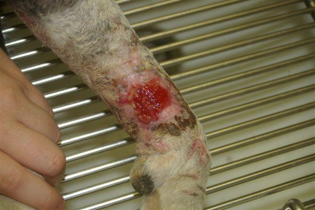
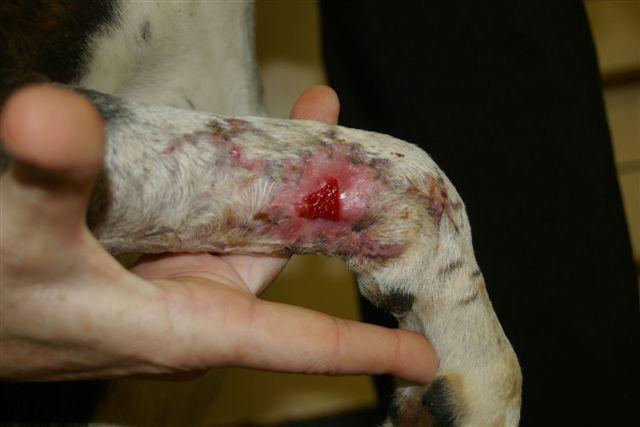
The wound is healing and granulating well. Antibiotics and pain killers are discontinued but the wound is bathed and re bandaged again on a weekly basis. Waggie is very happy and bright again.
Week Five:
The wound is contracting furthur and continuing to heal.
Week Six:
The wound is bathed and left open to continue healing.
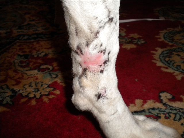
Week Nine:
The wound is basically completely healed. Pink scar tissue remains which will continue to contract until only a very small scar will be left. This shows how incredible the body is at healing itslef and regenerating tissue.
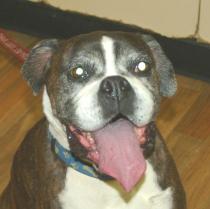
Waggie is her joyous self again! Even 8 months later there is no sign of recurrence of the tumour. Without the surgery to remove the tumour, Waggie would have succumbed to the disease and passed away by this time. Although there are no guarantees that the tumour will not come back….. for now Waggie is enjoying life to the fullest!